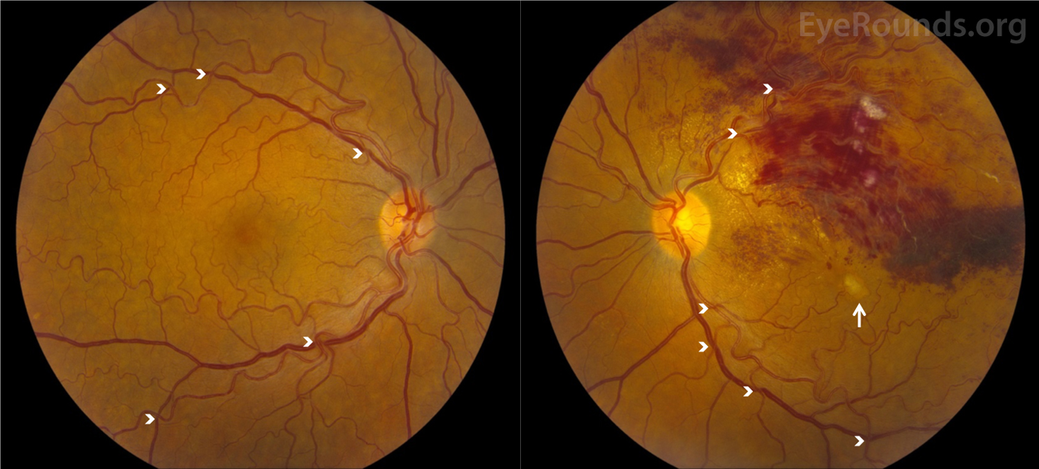
In addition, a further coordinate was recorded for those hemorrhages that were in the plane of the retina and/or optic nerve. The eye involved and the number and location of each hemorrhage were recorded relative to the optic nerve head in terms of clock hours and perceived plane, be that anterior to the optic nerve and/or retina, in the retinal nerve fiber layer, or finally occupying the subretinal space. We defined an ONH as being an isolated hemorrhage observed anywhere on or within 1 disc diameter of the optic nerve. The study received approval from the Human Research Ethics Committee at the Royal Australian and New Zealand College of Ophthalmologists. We conducted a prospective study of 35 consecutive patients presenting with symptoms of PVD of less than 2 months' duration who had a hemorrhage on or immediately adjacent to the optic nerve at presentation at a private multispecialty ophthalmic practice in Auckland, New Zealand. Furthermore, new technologies such as optical coherence tomography (OCT) have not been utilized to explore the patterns of vitreous separation that may influence the development of ONH, which is the aim of this study. The incidence of ONH in eyes that have recently undergone a PVD is estimated 7 to be 4% but, unlike disc hemorrhage associated with glaucoma, the clinical features in the setting of a PVD have not been well documented. 2,5 The benign nature of a disc hemorrhage in the presence of a PVD was highlighted by Hoyt et al 6 and more recently has been shown not to be a risk factor for the development of peripheral retinal breaks. 1-4 Equally, ONH has been documented in apparently healthy eyes, a subset being those subjects that have recently undergone a symptomatic posterior vitreous detachment (PVD). Optic nerve hemorrhages (ONH) are commonly associated with such optic neuropathies as primary open-angle glaucoma, ischemic optic neuropathy, and diabetic papillopathy. The clinical features of ONH together with OCT imaging may help to distinguish ONH associated with PVD from other hemorrhages found on or adjacent to the optic nerve.

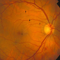
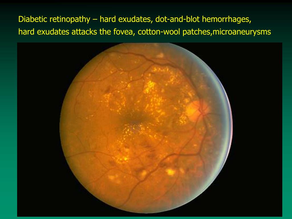
ONH hemorrhages associated with PVD are commonly found with persistent vitreopapillary adhesions as evidenced on OCT. One subretinal hemorrhage occurred in a patient with a central vitreopapillary adhesion. In all, 52 hemorrhages were identified, of which the majority were flame-shaped, although other types were seen including dot and blot hemorrhages. Three patterns of adhesion were identified: central, peripheral, and combined. Twenty of 30 eyes with ONH had evidence of persistent vitreopapillary adhesion. The number and location of each hemorrhage were recorded, together with relevant ophthalmic and demographic data.

#DOT BLOT HEMORRHAGES SERIES#
Methods:Ī consecutive series of patients presenting at a private multispecialty practice in Auckland, New Zealand, with symptoms of PVD with ONH underwent imaging of the optic nerve with digital retinal photography and OCT. Design:Ī prospective case series conducted at a private ophthalmic practice in Auckland, New Zealand.

To report and evaluate the clinical and optical coherence tomography (OCT) features of optic nerve hemorrhages (ONH) associated with spontaneous posterior vitreous detachment (PVD).


 0 kommentar(er)
